
You are petting your dog or cat and your hand passes over something that was not there before: a lump. Your mind races. The reality is that lumps and masses are extremely common in pets, especially as they get older, and many of them are completely benign. But there is no way to tell just by feel. A soft, squishy lump can be a harmless lipoma (fatty tumor) or something that needs attention. That is why diagnostic testing matters so much, and why we recommend testing rather than “watching and waiting” whenever a new mass appears.
At St. Petersburg Animal Hospital and Urgent Care, we perform fine needle aspirates (FNA, a quick needle-based cell collection) and surgical biopsies regularly, with transparent pricing so you know what to expect before we begin. We also use electrosurgery for mass removals, which provides precise tissue cutting with reduced bleeding. If you have found a lump on your pet, do not wait to have it checked. Call us at (727) 323-1311 or contact us to schedule an evaluation.
Why Looking and Feeling Is Never Enough
Signs of cancer in pets do not always look or feel a specific way. Experienced veterinarians cannot reliably distinguish a benign lipoma from a malignant soft tissue sarcoma by examination alone. Both can feel soft, both can be well-defined, and both can appear at the same sites. Surface characteristics are simply not diagnostic.
Cancer in pets is common. Roughly one in four dogs develops cancer at some point in their lifetime. Early diagnosis, before a tumor has grown significantly or spread, consistently correlates with better outcomes and more treatment options. The combination of earlier diagnosis and more options available is why we test early rather than observe.
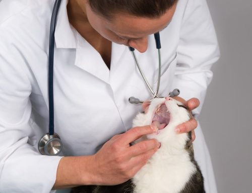
Fine Needle Aspiration: The Fastest First Step
How FNA Works
A fine needle biopsy uses a thin needle inserted into the mass to collect cells or fluid. The sample is placed on a slide, stained, and examined microscopically. The procedure takes only a few minutes and causes minimal discomfort. Most dogs and cats tolerate it with no sedation; it’s a little like getting a vaccine. Multiple areas of a mass or multiple masses can be sampled in a single visit.
Results are typically available within a few days from our reference laboratory. For some sample types, preliminary in-house assessment can begin immediately.
What FNA Can Tell Us
Cytology from FNA examines individual cells rather than tissue architecture. Skin cytology is particularly informative for superficial masses and skin lesions. When evaluating cytology, we’re looking for bacteria, fungal organisms, inflammatory cells, or cells that show signs of cancerous changes.
What cytology can reliably diagnose:
- Mast cell tumors: often exfoliate well and show distinctive granules visible under the microscope
- Lipomas: release fat droplets confirming their benign fatty tissue origin
- Cysts and infection: typically yield fluid containing specific cell types
- Reactive lymph nodes: distinguishes infection from lymphoma in many cases
- Round cell tumors: including histiocytomas, plasmacytomas, and transmissible venereal tumors
For any of these, a single FNA can provide a complete answer and guide the entire treatment plan.
When FNA Falls Short
Some tumor types do not release cells easily into a needle. Fibrosarcomas, poorly exfoliating mammary tumors, and certain other sarcomas produce samples where cells are sparse or unrecognizable as cancer even when the tumor is malignant. An FNA that returns as “reactive tissue” or “inadequate sample” does not rule out cancer.
A non-diagnostic FNA from a growing or suspicious mass should proceed to biopsy rather than result in monitoring. The limitations of cytology are real, and knowing when to move to the next test matters.
Biopsy: When Tissue Architecture Is Needed
How Biopsy Works
A biopsy removes a tissue sample rather than just cells. That tissue is sent for histopathologic examination by a board-certified veterinary pathologist, who examines the cellular arrangement, the relationship between cells, invasion patterns, and margin status.
Common biopsy types:
- Punch biopsy: a small circular cutting tool removes a cylinder of skin and underlying tissue; used for well-defined superficial masses
- Incisional biopsy: a wedge of tissue is removed from the mass without removing the whole thing; used for large or infiltrative masses where diagnosis before definitive surgery is important
- Excisional biopsy: the entire mass is removed and submitted for histopathology; combines treatment and diagnosis when the mass is small and accessible
- Needle core biopsy: a larger-gauge needle collects a core of tissue with preserved architecture; bridges FNA and surgical biopsy in some situations
Biopsies require sedation or anesthesia to ensure accurate sampling and patient comfort. Tumor diagnosis through histopathology provides information that cytology simply cannot.
What Histopathology Tells Us That FNA Cannot
Histopathology examines tissue architecture. This is where the most clinically important information lives:
- Tumor grade: how aggressive the cells look and how they are arranged, which predicts biological behavior
- Invasion patterns: whether the tumor is infiltrating surrounding structures
- Margin assessment: whether the removed tissue has clear margins (no tumor cells at the edge) or incomplete margins (tumor present at the cut edge, indicating a need for additional surgery)
- Definitive classification: the exact tumor type, which determines treatment, prognosis, and referral recommendations
Types of cancer that look identical on FNA can behave completely differently and require different treatments. Histopathology resolves that ambiguity.
Knowing whether a mass was completely excised with clear margins is one of the most practical outcomes of submitting any surgically removed mass. Without margin assessment, there is no way to know whether the surgery was curative.
Choosing Between FNA and Biopsy
The Clinical Decision
FNA is typically the first step because it is quick, low-cost, and low-risk. It answers the clinical question in many cases without anesthesia. Biopsy is recommended when:
- FNA returns non-diagnostic or inconclusive results from a suspicious mass
- The tumor type requires grading for treatment planning
- Margin assessment after removal is clinically important
- The mass is located where FNA yields poor results
- Clinical suspicion for malignancy is high despite a benign-appearing FNA
Mast cell tumors are among the most common skin tumors in dogs and tend to exfoliate well, making FNA both diagnostic and useful for staging. Grading, however, requires histopathology. A mast cell tumor diagnosed on FNA still needs biopsy after excision to determine grade and margin status.
Skin cancers range from completely benign (histiocytoma, which often resolves on its own) to highly malignant (squamous cell carcinoma, melanoma). FNA can distinguish them in many cases, but the definitive answer for margin-critical surgical planning requires histopathology.
About Electrosurgery for Mass Removal
We use electrosurgery during mass removals at St. Petersburg Animal Hospital. Electrosurgery uses controlled electrical current to cut tissue and cauterize bleeding vessels simultaneously, reducing blood loss, minimizing procedure time, and providing precise tissue cutting for masses in sensitive locations or near important structures. For pets undergoing mass removal, this translates to a cleaner procedure, faster closure, and a more efficient recovery.
What to Expect During and After Testing
The FNA Experience
Your pet is positioned comfortably. The needle is inserted and cells are collected over 30 to 60 seconds. Your pet goes home immediately. Results return within a few business days from our reference laboratory. Our team contacts you directly to walk through what the results mean and what the recommended next steps are.
The Biopsy Experience
Biopsies require sedation or general anesthesia. Pre-anesthetic bloodwork screens for metabolic concerns. The procedure takes 15 to 60 minutes depending on mass size and location. Your pet recovers at our facility and goes home the same day in most cases. An e-collar protects the incision site. Suture removal is scheduled for 10 to 14 days. Histopathology results return from the pathologist within one to two weeks.
Understanding Your Results
- Benign result: provides peace of mind and a monitoring plan. Some benign masses benefit from eventual removal; others require only periodic measurement.
- Malignant result: opens the conversation about grade, margins, additional testing, and treatment options including surgery, chemotherapy, radiation, or palliative care depending on the diagnosis.
- Inconclusive result: prompts discussion of whether biopsy is the next step and how the clinical picture should guide timing.
Our internal medicine and surgery services cover the diagnostic and treatment process. Our team explains findings in plain language, answers every question, and respects that the decision about next steps belongs to your family.

Frequently Asked Questions
Is FNA painful for my pet?
For most pets, FNA is well-tolerated without sedation. The needle is small, the procedure is brief, and many pets do not react at all. Topical anesthetic is available for sensitive locations.
Can I skip FNA and go straight to biopsy?
Yes, in some situations that is the right approach. For large, rapidly growing, or clinically suspicious masses, starting directly with excisional biopsy or incisional biopsy avoids the delay of an FNA that may not give a definitive answer anyway. Our team helps determine the most efficient path for your pet’s specific mass.
What if the FNA comes back normal but the mass keeps growing?
A growing mass that returns a benign or non-diagnostic FNA should not be dismissed. Repeat sampling or biopsy is appropriate. Growth is always meaningful clinical information.
How much does testing cost?
We publish our pricing transparently. Contact us to get current pricing before your appointment. There are no surprises.
A Lump Found Is a Lump That Can Be Evaluated
The hardest part of finding a lump is often the not knowing. At St. Petersburg Animal Hospital and Urgent Care, we move quickly from discovery to answers: same-day evaluation during our open hours, accessible pricing, and clear communication about what results mean and what comes next.
Contact us at (727) 323-1311 or visit us directly for a wellness exam or urgent care evaluation. Walk-ins are welcome for mass evaluations.

Leave A Comment